
Key parameters of the spectrograph:
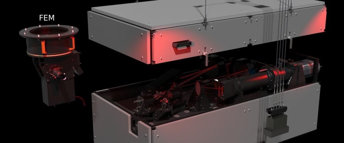 Three dimensional rendering of the spectrograph optomechanical design and of the front-end module (FEM) structural support assembly.
Full scientific descriptions:
Colby, J., et al. Design of radial velocity spectrograph for the Molėtai Observatory. Proc. SPIE 9147, Ground-based and Airborne Instrumentation for Astronomy (2014), http://dx.doi.org/10.1117/12.2056491
Colby, J., et al. Design and Construction of VUES: the Vilnius University Echelle Spectrograph, 2016, http://arxiv.org/abs/1601.06024
Additional information at the web page of Yale University: http://exoplanets.astro.yale.edu/instrumentation/mao.php
Construction of the spectrograph was financed from the European Structural and Investment Funds (contract No VP2-1.1-ŠMM-06-V-01-013).
Three dimensional rendering of the spectrograph optomechanical design and of the front-end module (FEM) structural support assembly.
Full scientific descriptions:
Colby, J., et al. Design of radial velocity spectrograph for the Molėtai Observatory. Proc. SPIE 9147, Ground-based and Airborne Instrumentation for Astronomy (2014), http://dx.doi.org/10.1117/12.2056491
Colby, J., et al. Design and Construction of VUES: the Vilnius University Echelle Spectrograph, 2016, http://arxiv.org/abs/1601.06024
Additional information at the web page of Yale University: http://exoplanets.astro.yale.edu/instrumentation/mao.php
Construction of the spectrograph was financed from the European Structural and Investment Funds (contract No VP2-1.1-ŠMM-06-V-01-013).
| Wavelength Range | λ = 400–900 nm |
| Spectral Resolution Modes, λ / δλ | 30000; 45000; 60000 |
| Echelle spectrum | 70 – 153 raws |
| Échelle Diffraction Grating | 31.6 grooves/mm |
| Instrumental Throughput | 25%, λ = 543 nm |
| Broad-spectrum optical fiber (FBPI) | fiber, Φ = 100 µm, l = 16 m |
| On-sky Fiber Aperture | 2.5 arcseconds |
| Spectrograph Detector | 4k x 4k x 15 μm pixel pitch |
| Temperature | -94oC |
Three dimensional rendering of the spectrograph optomechanical design and of the front-end module (FEM) structural support assembly.
Full scientific descriptions:
Colby, J., et al. Design of radial velocity spectrograph for the Molėtai Observatory. Proc. SPIE 9147, Ground-based and Airborne Instrumentation for Astronomy (2014), http://dx.doi.org/10.1117/12.2056491
Colby, J., et al. Design and Construction of VUES: the Vilnius University Echelle Spectrograph, 2016, http://arxiv.org/abs/1601.06024
Additional information at the web page of Yale University: http://exoplanets.astro.yale.edu/instrumentation/mao.php
Construction of the spectrograph was financed from the European Structural and Investment Funds (contract No VP2-1.1-ŠMM-06-V-01-013).
| CCD | E2V CCD47-10 |
| Array Size (pixels) | 1024 x 1024 |
| Pixel Size | 13 x 13 microns |
| Imaging Area | 13.3 x 13.3 mm (177 mm2) |
| Imaging Diagonal | 18.8 mm |
| Video Imager Size | 1.2” |
| Linear Full Well (typical) | 100K electrons |
| Dynamic Range | 83 dB |
| QE at 400 nm | 52% (MB); 75% (BB); 57% (UV) |
| Peak QE | 96% (MB); 86% (BB); 65% (UV) |
| Anti-blooming | none |


CCD photometer is equipped with filters of UPXYZVS photometric system.
The transmission curves of Vilnius photometric system
 Mean wavelength and half-widths of response functions
CCD camera for photometer:
Mean wavelength and half-widths of response functions
| U | P | X | Y | Z | V | S | |
| Mean wavelength (nm) | 345 | 374 | 405 | 466 | 516 | 544 | 656 |
| Half-width (nm) | 40 | 26 | 22 | 26 | 21 | 26 | 20 |
| CCD | E2V CCD47-10 |
| Array Size (pixels) | 1024 x 1024 |
| Pixel Size | 13 x 13 microns |
| Imaging Area | 13.3 x 13.3 mm (177 mm2) |
| Imaging Diagonal | 18.8 mm |
| Video Imager Size | 1.2” |
| Linear Full Well (typical) | 100K electrons |
| Dynamic Range | 83 dB |
| QE at 400 nm | 52% (MB); 75% (BB); 57% (UV) |
| Peak QE | 96% (MB); 86% (BB); 65% (UV) |
| Anti-blooming | none |
| Number of channels (CH1, CH2 and CH3) | 3 |
| Distance between the channels CH1 and CH2 | 26 mm to 50 mm (7 arcmin to 13 arcmin for F=13 m) |
| CH1 and CH3 | 20 mm (5,3 arcmin for F=13 m) |
| Possible deviation of CH3 | +/- 2 mm (31 arcs for F=13 m) |
| Number of filter wheels | 2 (for CH2 and for CH1+CH2) |
| Number of filters in each filter wheel | 10 in CH1 (5 pairs in CH1+CH3) |
| Diameter of filters | 13 mm |
| Number of aperture wheels | 2 (for CH2 and for CH1+CH3) |
| Number of apertures in each wheel | 10 in CH1 (5 pairs for CH1+CH3) |
| Diameter | 0,3 to 8,0 mm (5 arcs to 2 arcmin for F=13m) |
| Diameter of field eyepiece | 40 mm (10,2 arcmin for F=13 m) |
| Number of aperture microscopes | 2 (for CH2 and for CH1+CH3) |
| Magnification of aperture microscopes | 25X |
| Diameter of the field of microscopes | 8 mm (2 arcmin) |
| Number of Fabri lenses | 3 |
| Diameter of Fabri lenses | 14 mm |
| Focus length of Fabri lenses | 30 mm |
| Diameter of outcoming pupil | 2,5 mm |
| Spectral range | 300 to 650 nm (PMT - R647P) 185 to 850 nm (PMT - R1463P) |
| Temeperature of the PMTs | two stabilized temperatures -5°C and -15°C which can be set depending on the enviromental temperature |
| System of the PMT cooling | one stage thermoelectric cooler with actively ventilated heat absorber |
| Dimensions of the photometer | height 410 +/- 5 mm max diameter (including microscopes) 210 +/- 5 mm max diameter of the main frame 140 +/- 5 mm |
| Imaging area | 1340 x 1300 pixels |
| Pixel size | 20 x 20 μm |
| Linear area of CCD chip | 26.8 x 26.0 mm |
| Scientific grade back illuminated CCD chips with Unichron UV enhancenment coating | |
| Liquid Nitrogen cooling | |
| Spectral range | 300 - 1000 nm |
| 40 % at 300 nm | |
| > 90 QE at 500 - 650 nm | |
| Dark current | ≤ 1 ē/p/hr at -120C |
| Read noise at scan rate of 100 kHz | ≤ 5 ē |
| Full frame readout time | 18 s |
| Dynamic range | 16 bits |
| Positions filter wheel | 8 |
| Diameter of the filter | 50 mm |